Decoding Beck's Triad: Cardiac Tamponade for EMTs
Master Beck's Triad for the NREMT. Learn the pathophysiology of cardiac tamponade, how to identify the three classic clinical signs, and key field treatments.
· 14 min read
Read →
Learn how to recognize Cushing's Triad in the field, understand the Monro-Kellie doctrine, and differentiate increased intracranial pressure from systemic shock.
When you treat a severe head injury in the field, the clock moves differently. You are not just managing an obvious scalp laceration or a broken bone. You are fighting an invisible, high-pressure crisis locked completely inside a rigid bone vault. Early on, a patient with a traumatic brain injury might seem relatively stable. They might talk to you, follow basic physical commands, or simply complain of a massive headache. Beneath the surface, a lethal buildup of fluid or intracranial blood could be silently expanding.
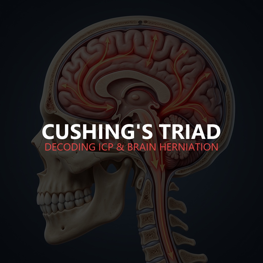
As an emergency provider, your absolute worst nightmare is missing the transition from a standard concussion to structural brain herniation. That is where understanding Cushing’s triad becomes your most vital NREMT prep asset. Named after pioneering American neurosurgeon Harvey Cushing, this clinical triad is a late, definitive distress flare that your patient’s brain is running out of room. This comprehensive guide will untangle the complicated pathophysiology of intracranial pressure, contrast head trauma vitals with standard shock, and give you a clear operational roadmap to protect your patient when every second counts.
To understand why Cushing’s triad happens, you have to look closely at the unique physics of the human head. This structural relationship is defined by a fundamental medical framework known as the Monro-Kellie doctrine.
Picture the skull as a completely unyielding, non-expandable solid box. Inside this closed container, three distinct physiological components exist in a delicate, balanced state:
This homeostatic balancing act works perfectly up to a specific mathematical tipping point. Once the bleeding or cerebral edema exceeds the body’s compliance limits, the internal pressure begins to climb sharply. Normal intracranial pressure ranges between 5 and 15 mmHg. When a traumatic mass effect pushes that number above 20 mmHg, the patient enters a state of critical intracranial hypertension. This structural bottleneck directly chokes off the blood flow entering the head, starving brain cells of oxygen.
When intracranial pressure rises to dangerous levels, the brainstem gets squeezed against the base of the skull. This triggers a frantic, multi-step survival reflex called the Cushing response. This reflex manifests as three highly specific changes in your patient’s vital signs.
As the pressure inside the skull approaches or exceeds the patient’s average blood pressure, the cerebral blood vessels get compressed flat. The brain instantly senses that it is suffocating from a lack of oxygen. In a desperate attempt to force blood past this obstruction, the sympathetic nervous system triggers a massive release of adrenaline.
This causes intense peripheral vasoconstriction, driving the systolic blood pressure up to extreme levels. However, the diastolic pressure often remains completely normal or drops slightly. This creates a massive gap between the two numbers, known clinically as a widening pulse pressure.
The sudden, massive spike in systemic blood pressure does not go unnoticed by the body’s internal regulatory systems. Specialized pressure sensors (baroreceptors) located inside the carotid arteries detect this intense surge of blood.
The brain attempts to protect the cardiovascular system by firing the parasympathetic nervous system via the vagus nerve, sending an immediate command to slow down. This causes the heart rate to drop down into a slow, bradycardic rhythm, often falling into the 40s or 50s. You are left with a patient who has a massively elevated blood pressure coupled with an abnormally slow pulse. (To review general cardiac function, see our guide on the cardiovascular system for EMTs).
The final component of the triad involves a direct physical assault on the respiratory center located inside the brainstem. As the mounting pressure compresses the medulla oblongata, the electrical signals that govern breathing become completely disrupted.
The patient’s breathing will change from a regular, smooth cadence to highly abnormal, unpredictable patterns. You will often spot Cheyne-Stokes respirations, which are characterized by deep, fast breaths followed by sudden, terrifying periods of total apnea. As the condition degrades further, this can transform into completely chaotic, agonal gasps.
One of the most dangerous traps for any student or rookie EMT is confusing the vital signs of systemic shock with those of a traumatic brain injury. Mixing these two up can lead to catastrophic treatment errors in the field.
To keep them straight in your mind, remember that increased intracranial pressure presents as the exact, structural opposite of hypovolemic shock. It is a complete vitals paradox. Differentiating increased intracranial pressure from systemic hypovolemic shock is crucial.
| Vital Sign Metric | Systemic Hypovolemic Shock | Critical Increased ICP (Cushing’s Triad) |
|---|---|---|
| Blood Pressure | Decreases significantly (Hypotension) | Increases significantly (Systolic Hypertension) |
| Pulse Pressure | Narrows or tightens | Widens significantly |
| Heart Rate | Accelerates rapidly (Tachycardia) | Slows down significantly (Bradycardia) |
| Respiratory Rate | Fast, shallow, and rapid (Tachypnea) | Irregular, abnormal, or deep (Cheyne-Stokes) |
| Skin Presentation | Pale, cold, clammy, and mottled | Typically normal, dry, or flushed |
In a typical multi-trauma scenario, a patient might have a severe head injury along with internal bleeding in their abdomen. If you notice their heart rate is climbing while their blood pressure is dropping, the body is fighting systemic blood loss, not head trauma. If you see their blood pressure climbing to 190 while their heart rate drops to 48, the brain is actively suffocating under massive internal pressure. To see how these neurological signs contrast with the mechanical cardiovascular pressure of a squeezing pericardial sac, check out our guide on Beck’s Triad.
Let us be entirely candid about what Cushing’s triad actually means when you spot it on a call. This is not an early, subtle warning sign. It is a late, dramatic distress flare.
When all three components of the triad are visible, your patient has already reached the absolute edge of a terminal physiological cliff. The pressure inside the skull has become so overwhelming that the brain tissue itself is being physically forced downward out of its natural position.
This catastrophic shifting is known as brain herniation. As the uncus or tonsils of the brain get driven down through the narrow opening (foramen magnum) at the base of the skull, the brainstem gets crushed. This leads to irreversible neurological destruction, followed rapidly by complete respiratory and cardiac arrest if immediate surgical intervention is delayed. Patients showing these signs carry an exceptionally high mortality rate.
When you identify Cushing’s triad during your patient assessment, your primary goal is to slow down the herniation process and buy the patient enough time to reach a neurosurgeon. Prehospital management relies on precise, calculated interventions that optimize cerebral perfusion while keeping intracranial pressures as low as possible.
The three hallmark components are systolic hypertension with a widening pulse pressure, bradycardia, and irregular, decreased respirations. These physical shifts reflect the nervous system’s terminal response to dangerous elevations of intracranial pressure inside the skull.
Cushing’s triad is an exceptionally late sign of increasing intracranial pressure. When these three vital sign changes appear together, it means the body’s natural compensatory systems have failed completely, and brain herniation is actively imminent.
The two conditions present with completely opposite vital signs. Systemic hypovolemic shock displays low blood pressure, a rapid heart rate, and fast, shallow breathing. Conversely, critical increased intracranial pressure shows high blood pressure, a slow heart rate, and irregular, slow respirations.
A widening pulse pressure occurs when the gap between the systolic blood pressure and the diastolic blood pressure grows significantly larger. In head injuries, the sympathetic nervous system drives the upper systolic number up to extreme levels while the lower diastolic number stays normal or drops.
The heart rate slows down due to a powerful parasympathetic nervous system response triggered by the vagus nerve. When the body drives up its blood pressure to push oxygen to the brain, internal sensors detect the massive pressure surge and attempt to slow down the heart to protect the vascular system.
Test your understanding of intracranial pressure dynamics, Monroe-Kellie physics, and head trauma assessments with this interactive 5-question NREMT-style quiz.
You are treating a 42-year-old female who fell down a flight of concrete stairs. She is unresponsive to verbal commands and withdraws from painful stimuli. Her vital signs are BP 178/62, HR 48, RR 8 and irregular. What is the primary physiological driver of her slow heart rate?
For a deeper look at how these life-threatening clinical signs present across different emergency medical runs, you can review this helpful breakdown on how to Understand Cushing’s Triad with Three Patient Scenarios. This visual scenario analysis walks you through real-world medical situations, explicitly showing how to contrast traumatic brain injuries from systemic blood loss in real time.
Filed Under
About the Author
Veteran EMT with 13+ years of field experience in EMS. I built EMT Training Station to give aspiring first responders the honest, practical information I wish I'd had when starting out — covering training, certification, gear, and career advancement.
Master Beck's Triad for the NREMT. Learn the pathophysiology of cardiac tamponade, how to identify the three classic clinical signs, and key field treatments.
Master the Rule of Nines for the NREMT. Learn burn classifications, adult vs. pediatric calculations, fluid resuscitation formulas, and critical field care.
Learn how to write professional patient care report (PCR) narratives that protect your EMS career. Master SOAP vs CHART models and document critical negatives.