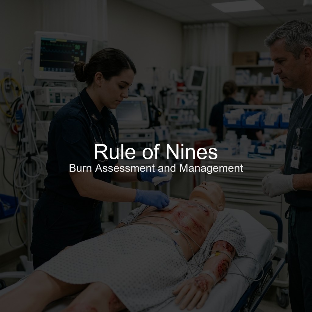
Rule of Nines for EMTs: Burn Assessment and Management
Master the Rule of Nines for the NREMT. Learn burn classifications, adult vs. pediatric calculations, fluid resuscitation formulas, and critical field care.
· 12 min read
Read →
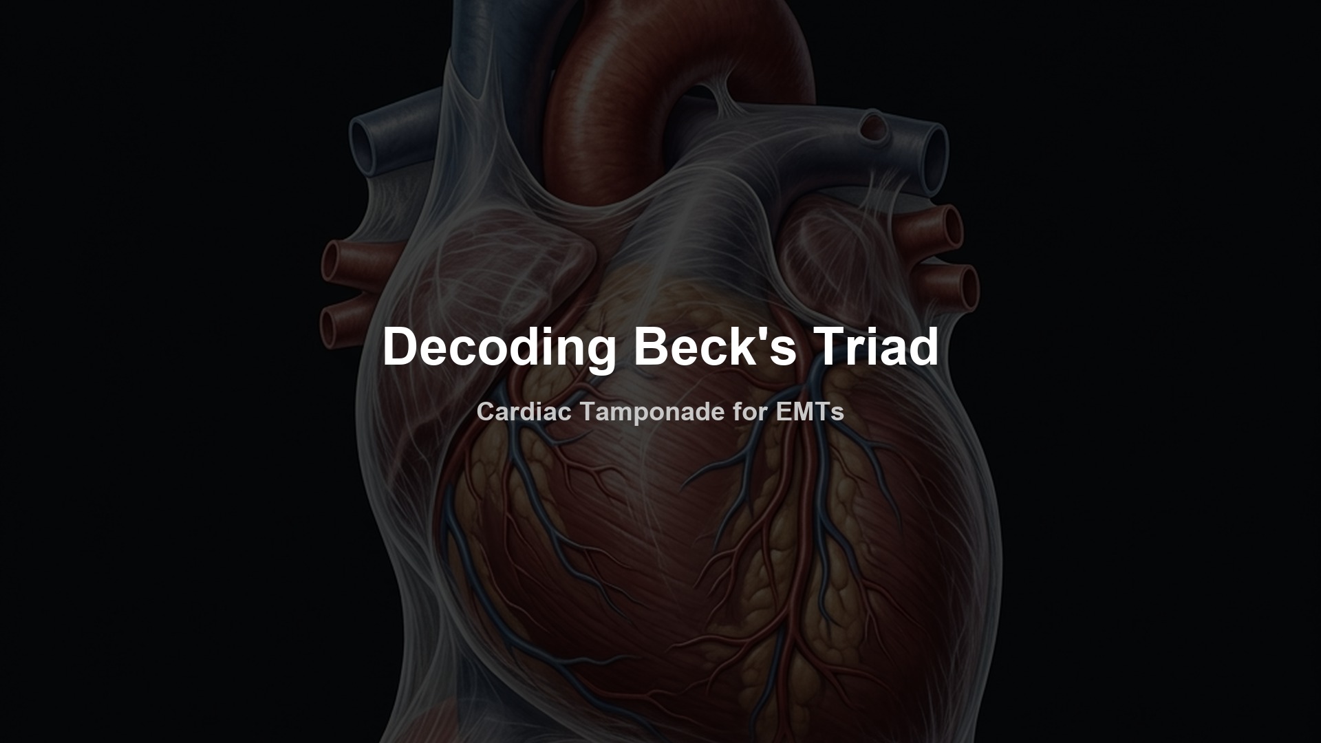
Master Beck's Triad for the NREMT. Learn the pathophysiology of cardiac tamponade, how to identify the three classic clinical signs, and key field treatments.
When you respond to a high-velocity motor vehicle collision or a penetrating chest wound, your mind immediately runs through a checklist of life-threatening thoracic injuries. You check for a sucking chest wound, a tension pneumothorax, and flail segments. However, there is another silent killer hiding within the chest cavity that can stop the heart from pumping entirely.
That killer is cardiac tamponade. As a student preparing to pass the NREMT exam or a field EMT facing a critical trauma patient, recognizing the signs of tamponade quickly is vital. The standard key to unlocking this diagnosis is a clinical triad first described by surgeon Claude Beck in 1935.
To master Beck’s Triad, you must understand the underlying pathophysiology. It is not enough to simply memorize three symptoms. You need to know exactly why the heart fails under this mechanical pressure, how to distinguish this cardiovascular crisis from a brain injury, and how to manage the patient in the field.
When your patient is in critical condition, you do not have time to second-guess your assessment. For the NREMT exam and real-world emergencies, you can use the 3 Ds mnemonic to recall the three clinical signs of Beck’s Triad:
| Triad Component | Physiological Explanation | How to Assess |
|---|---|---|
| Distant Heart Sounds | Fluid surrounds the heart, acting as an acoustic barrier. | Auscultate the apical pulse directly over the chest wall. |
| Distended Neck Veins | Backpressure of blood in the superior vena cava. | Inspect the jugular veins with the patient sitting at a 45-degree angle. |
| Decreased Blood Pressure | Compressed ventricles cannot maintain stroke volume. | Measure the blood pressure, noting a narrowing pulse pressure. |
To understand Beck’s Triad, you must first understand the anatomy of the heart and its protective casing. (For a quick refresher, review our guide on the cardiovascular system for EMTs). The heart is nested within a tough, fibrous sac called the pericardium.
Under normal conditions, a tiny amount of lubricating fluid (about 15 to 50 milliliters) sits between the heart muscle and the pericardial sac. This fluid allows the heart to beat smoothly without friction.
The pericardium is strong and inelastic. It does not stretch easily. When trauma causes blood or fluid to leak into this space, the pericardial sac quickly fills to capacity. Once the space is full, any additional fluid exerts direct pressure on the heart muscle. This condition is known as cardiac tamponade.
Think of the pericardium as a tight leather jacket. If you try to inflate a balloon inside a tightly zipped leather jacket, the balloon cannot expand. Similarly, as fluid builds up inside the inelastic pericardium, it squeezes the chambers of the heart.
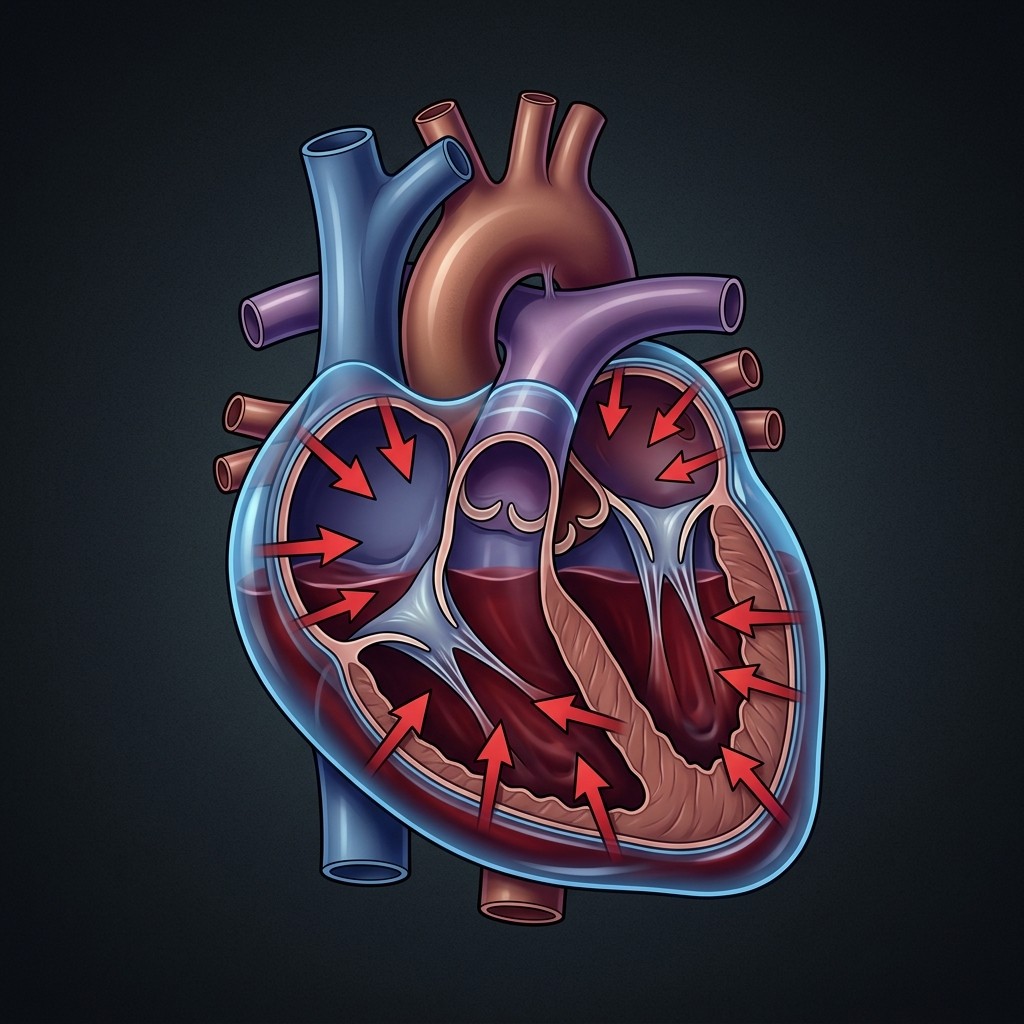
This compression directly impacts the heart’s ability to fill with blood during diastole. The right side of the heart has thin walls and operates under low pressure, making it the first area to collapse under the external squeeze.
Because the right ventricle cannot expand, it cannot receive blood returning from the body. This creates a severe drop in preload. Since the heart cannot fill, it cannot pump blood forward to the lungs and the left ventricle.
This drop in stroke volume leads directly to a crash in cardiac output. This is a classic form of obstructive shock. The plumbing is intact, the pump wants to work, but a mechanical barrier prevents it from filling and contracting. (To study the different types of shock in detail, review our comprehensive guide on shock for EMTs).
Let’s look closely at the three components of Beck’s Triad and how the underlying pathophysiology creates them.
When you place your stethoscope on the patient’s chest, the normal “lub-dub” sounds of the tricuspid, mitral, aortic, and pulmonary valves closing should be clear. In cardiac tamponade, fluid accumulates around the heart, creating an acoustic barrier.
Instead of hearing the crisp snapping of the heart valves, the sound waves must travel through a layer of blood or fluid before reaching your stethoscope. This makes the heart sounds seem distant, quiet, or muffled.
Auscultate the heart sounds early in your assessment if you suspect thoracic trauma. Compare what you hear to the lung sounds. If the lung sounds are clear and equal but the heart sounds are barely audible, this points directly to pericardial fluid accumulation.
As the pericardial fluid squeezes the right atrium and ventricle, the pressure inside the right side of the heart rises. The blood returning from the upper body via the superior vena cava cannot flow into the collapsed right atrium.
This creates a massive traffic jam in the venous system. Blood backs up into the large veins of the neck, leading to prominent jugular venous distention.
You should assess for JVD by placing the patient in a semi-Fowler’s position (45-degree angle). If the neck veins remain bulging and visible in this position, it indicates elevated central venous pressure.
As the heart’s chambers are compressed, stroke volume (the amount of blood pumped per beat) drops. The body tries to compensate by increasing the heart rate (tachycardia) and constricting blood vessels.
Eventually, these compensatory mechanisms fail. The blood pressure drops, resulting in systemic hypotension.
In addition to hypotension, you will often notice a narrowing pulse pressure. The pulse pressure is the difference between the systolic and diastolic blood pressure. In cardiac tamponade, the systolic pressure drops because the heart cannot pump blood out. Meanwhile, the diastolic pressure remains stable or rises slightly due to systemic vasoconstriction. If you see a blood pressure of 90/74, the pulse pressure is only 16 mmHg, which is a major warning sign.
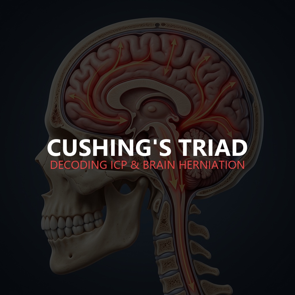
A common trap for NREMT students is confusing Beck’s Triad with Cushing’s Triad. Both are critical trauma indicators, but they represent entirely different physiological systems and emergencies.
| Feature | Beck’s Triad | Cushing’s Triad |
|---|---|---|
| Primary Condition | Cardiac Tamponade (Cardiovascular) | Increased Intracranial Pressure (Neurological) |
| Organ Affected | Heart (Pericardial Sac) | Brain (Intracranial Vault) |
| Blood Pressure | Hypotension (Narrowing Pulse Pressure) | Hypertension (Widening Pulse Pressure) |
| Heart Rate | Tachycardia (Rapid Heart Rate) | Bradycardia (Slow Heart Rate) |
| Respirations | Rapid, shallow breathing (Tachypnea) | Irregular, cheyne-stokes respirations |
| Key Signs | JVD, Muffled Heart Sounds | Bradycardia, Hypertension, Irregular Breathing |
Understanding this difference is crucial for your exams. Beck’s Triad is a cardiovascular failure caused by fluid squeezing the heart. Cushing’s Triad is a neurological failure caused by rising pressure inside the skull, which triggers the brainstem to slow the heart rate and drive up systolic blood pressure to force blood into the brain. (You can read more about intracranial pressure in our guide on neurological assessment for EMTs and our post on decoding Cushing’s Triad).
In the prehospital environment, cardiac tamponade is an extreme emergency that requires rapid transport. The definitive treatment for tamponade is pericardiocentesis, a procedure where a physician inserts a needle into the pericardial sac to drain the fluid. This is far outside the scope of practice for EMTs and paramedics in the field.
Your focus must be on rapid identification, stabilization, and immediate transport to a trauma center:
Beck’s Triad is one of the most critical clinical concepts you need to know. Remember the 3 Ds (Distant heart sounds, Distended neck veins, and Decreased blood pressure) and connect them directly to the mechanical squeeze of cardiac tamponade.
When preparing for your NREMT exam, practice identifying this triad in clinical scenarios. Look for clues like penetrating chest trauma (such as a stab wound to the chest) followed by JVD and muffled heart sounds. By understanding the underlying anatomy and pathophysiology, you will be ready to make the right call on the exam and save a life in the field.
Take this interactive, 5-question NREMT-style practice quiz to test your understanding of Beck’s Triad, cardiac tamponade, and shock management.
You respond to a local bar where a 28-year-old male has been stabbed in the chest. The patient is alert but restless, complaining of severe shortness of breath. Vital signs are BP 92/76, HR 118, RR 22, and SpO2 91% on room air. Neck veins are noticeably bulging, and heart sounds are quiet and difficult to hear. What is the most likely diagnosis?
The three components of Beck’s Triad are hypotension (decreased blood pressure), jugular venous distention (JVD or distended neck veins), and muffled (distant) heart sounds.
Beck’s Triad indicates cardiac tamponade, which is a cardiovascular emergency where fluid accumulates in the pericardial sac. Cushing’s Triad indicates increased intracranial pressure (ICP), which is a neurological emergency characterized by bradycardia, irregular respirations, and widening pulse pressure.
Fluid accumulating in the pericardial sac puts pressure on the heart muscle. This pressure prevents the ventricles from fully expanding and filling with blood, drastically reducing the stroke volume and cardiac output, leading to obstructive shock.
Filed Under
About the Author
Veteran EMT with 13+ years of field experience in EMS. I built EMT Training Station to give aspiring first responders the honest, practical information I wish I'd had when starting out — covering training, certification, gear, and career advancement.
Master the Rule of Nines for the NREMT. Learn burn classifications, adult vs. pediatric calculations, fluid resuscitation formulas, and critical field care.
Learn how to write professional patient care report (PCR) narratives that protect your EMS career. Master SOAP vs CHART models and document critical negatives.
Learn how to recognize Cushing's Triad in the field, understand the Monro-Kellie doctrine, and differentiate increased intracranial pressure from systemic shock.