Decoding Beck's Triad: Cardiac Tamponade for EMTs
Master Beck's Triad for the NREMT. Learn the pathophysiology of cardiac tamponade, how to identify the three classic clinical signs, and key field treatments.
· 14 min read
Read →
A complete EMT guide to the cardiovascular system: heart anatomy, electrical conduction, cardiac emergencies, and prehospital interventions.
Welcome to an in-depth exploration of the cardiovascular system and its significance in the field of emergency medical services. As an Emergency Medical Technician (EMT), your knowledge of this intricate system, the conditions that can arise, and the interventions necessary to save lives is fundamental to your role.
This comprehensive guide titled ‘The Cardiovascular System: EMT Interventions for Heart Conditions’ is designed to deepen your understanding and equip you with the practical knowledge you need in your life-saving work. In this five-part series, we will journey through the intricacies of the cardiovascular system, delve into the most common heart conditions you’ll encounter as an EMT, review essential interventions, and underline the importance of patient education in preventing cardiovascular disease.
Whether you’re a seasoned veteran in emergency medical services or an aspiring EMT just starting your career, this guide offers invaluable insights that will enhance your skills and enrich your professional journey. Let’s dive in and unravel the complexities of the cardiovascular system and the crucial role you play as an EMT in managing heart conditions.
The cardiovascular system, also called the circulatory system, is a complex biological network necessary for life. The cardiovascular system, which includes the heart, blood vessels, and blood, works tirelessly to supply oxygen and nutrients to all body cells and remove waste.
The heart is located centrally in the body. The heart is a relatively small muscle—about the size of a fist—but it has the enormous task of distributing blood throughout the body. The heart’s four chambers (two atriums and two ventricles) coordinate their contractions and relaxations to keep the heart beating at a steady rate. Blood is pumped out of the heart by the ventricles after being received by the atria. As a result of this cycle, blood is either oxygenated in the lungs (via the pulmonary circuit) or transported throughout the body (via the systemic circuit).
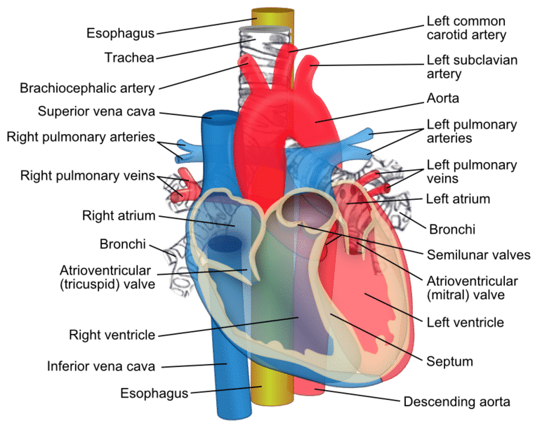
Take a moment to get to know the following anatomical structures of the heart as well as their functions
An EMT’s knowledge of the intricate electrical conduction system of the heart is indispensable. The heartbeat is coordinated by electrical impulses generated by this system, which also triggers the heart’s contractions. Any disturbance in this system can lead to arrhythmias, or irregular heart rhythms, which can be life-threatening if not treated immediately. A significant part of this system consists of two main nodes: the Sinoatrial (SA) node and the Atrioventricular (AV) node. Let’s explore these nodes and the heart rates typically associated with each.
The Sinoatrial node, or SA node, is often termed as the heart’s natural pacemaker. Located in the right atrium, it is responsible for initiating each heartbeat. The SA node generates electrical impulses that spread throughout the atria, causing them to contract and pump blood into the ventricles.
In a healthy adult at rest, the SA node fires between 60 to 100 times per minute. This is considered a normal resting heart rate. However, factors such as fitness level, emotional state, medication use, and age can influence this rate.
Situated in the lower part of the right atrium, the Atrioventricular or AV node serves as a critical gateway in the heart’s electrical conduction system. It receives the electrical impulses from the SA node, delays them slightly to allow the ventricles to fill with blood, and then transmits them to the ventricles causing them to contract.
In the event of an SA node malfunction, the AV node can initiate electrical impulses on its own, but at a slower rate—usually between 40 and 60 beats per minute. This slower rhythm is often referred to as a junctional rhythm.
Once the electrical impulse leaves the AV node, it travels along the Bundle of His and then the Purkinje fibers, causing the ventricles to contract. If both the SA and AV nodes fail, these structures can also generate impulses, albeit at an even slower rate—around 20 to 40 beats per minute.

The cardiac cycle is an intricate sequence of events that takes place in the heart during one complete heartbeat. This vital process, occurring approximately 60 to 100 times per minute in a healthy resting adult, involves a series of coordinated contractions and relaxations of the heart chambers to pump blood throughout the body.
The cardiac cycle can be divided into two main phases: systole and diastole.
Systole is the phase during which the heart contracts. It begins with the closure of the atrioventricular valves (tricuspid and mitral valves) to prevent backflow into the atria. This closure results in the first heart sound, often described as “lub.”
This phase is divided into two stages:
Diastole is the phase during which the heart relaxes and fills with blood. It begins with the closure of the semilunar valves, preventing backflow into the ventricles, resulting in the second heart sound often described as “dub.”
This phase is divided into three stages:
This systematic and rhythmical contraction and relaxation process of the cardiac cycle ensures the effective circulation of blood, providing oxygen and nutrients to the body’s tissues and organs.
Blood is transported throughout the body via an extensive and complex network of arteries, veins, and capillaries. Each is constructed and used in a different way. The oxygenated blood leaves the heart through the arteries, which are strong and durable. The veins carry the blood that has been deprived of oxygen back to the heart through one-way valves. Capillaries are the tiniest and most numerous blood vessels, and they play a key role in transporting oxygen, nutrients, and waste products from the circulatory system to the body’s tissues.
Blood is a unique bodily fluid that serves many important purposes. It’s crucial to our immune response and helps regulate temperature and waste removal, among other functions.
There are two primary reasons why EMTs need to have a solid understanding of the cardiovascular system. Heart-related emergencies occur frequently, to begin with. Intense chest pain, shortness of breath, and mental confusion are all symptoms that should alert a person to the possibility of cardiovascular disease. An EMT’s ability to recognize these symptoms and to interpret their potential meanings is critical because it informs their decisions and actions in the field.
Second, EMTs are frequently the first healthcare providers to engage with a patient in the “golden hour,” the period immediately following an emergency in which interventions can have the greatest possible effect on the patient’s outcome. This is especially true for those who suffer from cardiovascular disease. In the case of a heart attack, for instance, the damage can be permanent in a matter of minutes. If a patient receives a diagnosis and begins treatment quickly, their chances of survival and recovery are greatly increased.
Therefore, it’s not just helpful to have a good grasp of the cardiovascular system’s operations; it’s necessary. It lays the groundwork for identifying and handling many of the most common emergencies an EMT will encounter on the job. A healthcare provider’s ability to correctly diagnose a patient’s condition, begin effective treatment, and communicate this information to colleagues can literally be a matter of life and death.
A wide variety of cardiovascular conditions are common among the patients EMTs see in their role as first responders. Understanding the signs and symptoms of these diseases is essential in order to provide appropriate treatment in a timely manner. Heart attacks, arrhythmias, heart failure, and hypertension are just some of the most common cardiovascular emergencies seen by EMTs, and they will all be discussed in this section.
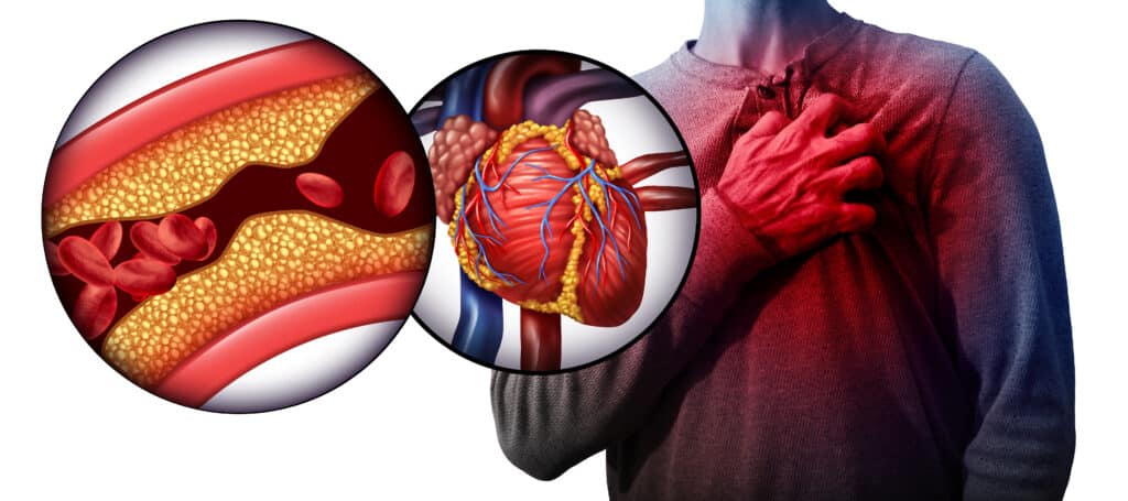
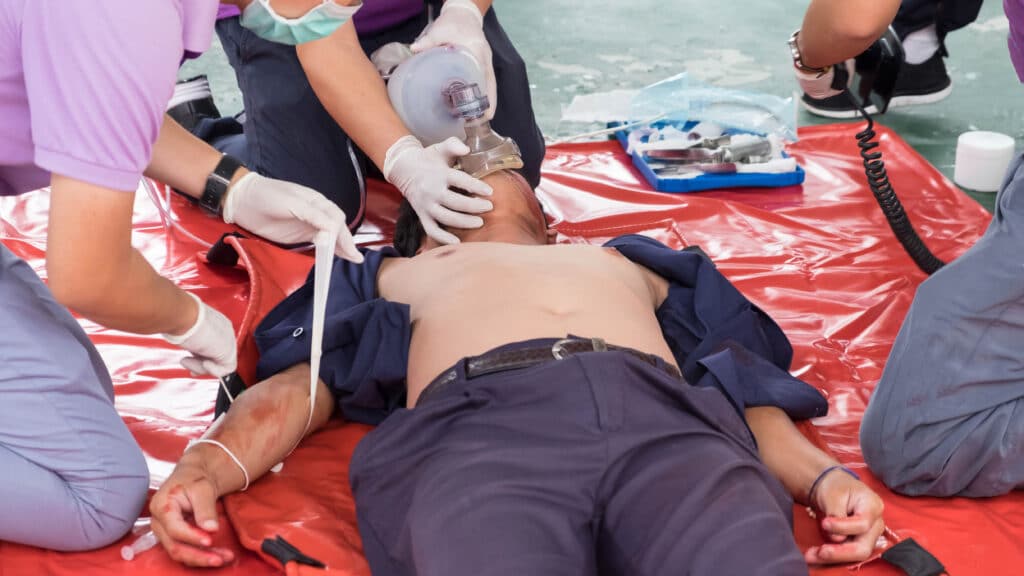
A heart attack, or myocardial infarction, is a life-threatening emergency that requires immediate medical attention. As an EMT Basic, your primary focus is to recognize the signs and symptoms of a heart attack and provide prehospital care to stabilize the patient while transporting them to the nearest appropriate medical facility. Let’s review the steps an EMT Basic can take to manage a suspected heart attack, including the use of oxygen, nitroglycerin, and baby aspirin.
Recognizing a heart attack can be challenging, as symptoms can vary greatly among individuals. Classic symptoms include chest pain, shortness of breath, nausea, vomiting, sweating, and discomfort radiating to the jaw, neck, or arms. Women, in particular, may present with less typical symptoms, such as fatigue, dizziness, or abdominal discomfort. Always consider a heart attack in any patient presenting with chest discomfort or other signs suggestive of heart disease.
Initially, aim to comfort and reassure the patient. Minimize their physical activity and help them into a position that eases their discomfort—usually a semi-sitting position.
Oxygen Administration
Administer supplemental oxygen only if the patient is dyspneic, hypoxemic (SpO2 < 94%), or shows signs of heart failure or shock. Routine administration of high-flow oxygen to non-hypoxic MI patients is no longer recommended under modern guidelines as hyperoxia causes coronary vasoconstriction and may increase myocardial injury.
Nitroglycerin Administration
If the patient has a prescription for nitroglycerin, and their blood pressure is within a safe range (usually systolic blood pressure above 90-100 mmHg), assist them in taking their medication. Nitroglycerin can help to dilate coronary arteries and improve blood flow to the heart muscle.
Always confirm with medical control or follow your local protocols before assisting with nitroglycerin administration, as there are contraindications, such as recent use of erectile dysfunction medications, to be aware of.
Baby Aspirin Administration
Chewable baby aspirin (typically four 81 mg tablets for a total of 324 mg) is often given to heart attack patients. Aspirin acts as an antiplatelet agent, reducing clot formation and helping to restore blood flow in the blocked artery.
Always ask about an allergy to aspirin or a history of gastrointestinal bleeding before administration and make sure the patient chews the aspirin for faster absorption. Again, follow your local protocols or obtain medical control clearance before administering aspirin.
Cardiac Monitoring and Rapid Transport
Monitor the patient’s vital signs closely and prepare for rapid transport to the hospital. If available and allowed by protocol, perform a 12-lead ECG. Time is crucial in a heart attack—every minute counts.
While EMT-Basics may not perform invasive procedures, their role in early recognition, initial management, and rapid transport of heart attack patients can have a significant impact on patient outcomes. Providing oxygen, assisting with nitroglycerin and aspirin administration, and facilitating rapid transport are all critical steps in the chain of survival for heart attack patients.
Arrhythmias represent abnormalities in the heart’s rhythm, triggered by issues within the heart’s electrical conduction system. Arrhythmias vary widely, from benign to severe. Symptoms can range from palpitations and fatigue to loss of consciousness or even cardiac arrest in extreme cases. Medication or implantable devices may be necessary for treatment, depending on the type and severity of the arrhythmia.
Heart failure is a chronic condition where the heart can’t pump sufficient blood to meet the body’s demands. Tiredness, shortness of breath, swelling in the legs, and a racing heart could be symptoms. Although there is currently no cure for heart failure, it can often be controlled with medication and behavioral modifications.
Hypertension is when the blood’s force against artery walls is consistently too high. Untreated hypertension is associated with serious outcomes such as heart attack, stroke, and kidney disease, despite the fact that the condition is often asymptomatic in its early stages. Treatment focuses on controlling blood pressure through lifestyle modifications and medication.
The ability of EMTs to detect these heart conditions early on can have a significant effect on patient outcomes. For instance, recognizing the symptoms of a heart attack and starting treatment before getting to the hospital can reduce the amount of damage done to the heart and increase the chances of survival.
EMTs can better anticipate and manage emergencies by having a firm grasp on the potential complications associated with these conditions. Cardiac arrest can be the result of a heart attack or certain types of arrhythmia, making prompt EMT intervention crucial.
When you work as an EMT, you do so as part of a larger medical group. The patient will receive the best possible follow-up care and treatment if the emergency room and other healthcare providers have access to accurate and timely information.
Emergency Medical Technicians (EMTs) are often the first healthcare professionals on the scene of a medical emergency. Assessing and diagnosing cardiovascular conditions quickly and accurately can have a major effect on patient outcomes. The electrocardiogram (ECG) is one example of a diagnostic tool that is used by paramedic level providers. As an EMT you will need to rely on your patient assessment skill and ability to identify key symptoms in order to determine if your patient is having a cardiac emergency.
An essential part of the EMT’s evaluation is the initial conversation with the patient. During this exchange, EMTs can ask the patient and any witnesses present for crucial details about their condition. First, EMTs must determine the patient’s “chief complaint,” or the primary ailment that prompted them to call for help. Symptoms of a heart problem may include discomfort in the chest, difficulty breathing, dizziness, and fatigue.
Next, EMTs investigate the patient’s history of the present illness (HPI), which is a thorough description of the onset and evolution of the symptoms. Performing such an investigation can help doctors better understand the patient’s condition and its severity. Structured tools like SAMPLE history and OPQRST provide EMTs with a reliable framework for this patient interview.
The patient’s medical history is also recorded by the EMT, with special focus on the presence or absence of cardiovascular disease. The last thing the patient ingested orally (whether it was food, liquid, or medication) can help the EMT predict potential complications and determine the best course of treatment.
The EMT’s ultimate goal is to determine what precipitated the current illness. Consider the patient’s activity level, mental health, environmental stresses, and any other relevant factors. The EMT can gain valuable insight from this background information as they investigate potential causes of the symptoms.
Once the EMT has finished talking to the patient, they will perform a physical examination. In the first phase of this checkup, the examiner will form an overall impression of the patient based on their demeanor, gait, and skin condition. All abnormalities that may indicate a problem will be investigated by EMTs.
The following important indicators are blood pressure, heart rate, respiratory rate, and oxygen saturation. Each of these readings has the potential to shed light on the patient’s cardiovascular health and reveal any impending crises.
The next step is a more in-depth physical examination, during which the doctor may use a stethoscope to listen to the patient’s heart and lung sounds, look for signs of poor circulation on the skin (such as pallor, coolness, or bluish tint), and check for swelling in the legs and ankles, which may indicate heart failure.
The EMT can then formulate a primary impression based on the collected data. The EMT’s initial assessment of the patient’s symptoms serves as a starting point for determining the most appropriate course of treatment and can be revised as new information becomes available.
Transferring the patient to the hospital marks the completion of the process. The EMT gives a handover, which is a report detailing their findings and the care they gave the patient in a clear and concise format. The continued success of the hospital’s care delivery depends on this data.
In conclusion, the successful management of cardiovascular conditions relies heavily on thorough evaluation and correct diagnosis. The ability of EMTs to quickly gather and interpret information is crucial, as it will direct their interventions and potentially save the patient’s life.
Test your understanding of heart anatomy, the cardiac cycle, and field management of cardiac emergencies with this interactive 5-question NREMT-style quiz.
You are assessing a patient complaining of crushing chest pain. If you suspect an acute myocardial infarction, which of the following is the most appropriate dosage and administration of aspirin for an EMT to provide?
The most common cardiac emergencies EMTs encounter are heart attacks (myocardial infarctions), arrhythmias, heart failure, and hypertensive crises. Recognizing the signs and symptoms of each is critical because early intervention in the field directly affects patient outcomes.
An EMT Basic can administer supplemental oxygen if the patient is hypoxemic (SpO2 < 94%) or in respiratory distress, assist the patient with prescribed nitroglycerin if blood pressure allows, administer chewable aspirin (324 mg), perform cardiac monitoring if available, and initiate rapid transport to the nearest appropriate facility. Every minute without reperfusion increases heart muscle damage.
The sinoatrial (SA) node is the heart’s natural pacemaker, located in the right atrium. It generates the electrical impulses that trigger each heartbeat at 60–100 bpm. Failure of the SA node can cause dangerous bradycardias or arrhythmias — conditions EMTs must recognize and manage in the field.
Filed Under
About the Author
Veteran EMT with 13+ years of field experience in EMS. I built EMT Training Station to give aspiring first responders the honest, practical information I wish I'd had when starting out — covering training, certification, gear, and career advancement.
Master Beck's Triad for the NREMT. Learn the pathophysiology of cardiac tamponade, how to identify the three classic clinical signs, and key field treatments.
Master the Rule of Nines for the NREMT. Learn burn classifications, adult vs. pediatric calculations, fluid resuscitation formulas, and critical field care.
Learn how to write professional patient care report (PCR) narratives that protect your EMS career. Master SOAP vs CHART models and document critical negatives.