How Does the Heart Work?

The purpose of studying the anatomy and physiology of the heart is to understand how this natural pumping mechanism works. Below are notes that supplement your class or home study program. Given the importance of anatomy and physiology in paramedicine it is recommended that students use additional easy to understand study material.
As an EMT or Paramedic, the heart’s health and function are among the most important aspects of Emergency Medicine. It is one of a triad of life providing aspects of the human body. The other two being a person’s airway and ability to breath. In this post, we will explore the anatomical structure of the heart.
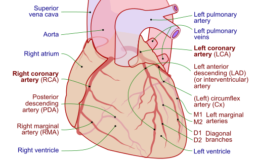
You will notice a diagram to the left. This lays out all of the basic structures of the heart that the emergency medical professional needs to concern themselves with. In a later post, we will explore the physiology (function) of the heart.
The Path Blood Takes Through The Heart:
Blood enters the heart through the superior and inferior vena cava. From there it enters the right atrium where it is pushed through the atrioventricular (tricuspid) valve into the right ventricle. From the right ventricle, the blood is pushed through the pulmonic (semilunar) valve into the pulmonary arteries (which are the only arteries in the body that carries deoxygenated blood).
After exchanging gases in the lungs, the now oxygenated blood reenters the heart through the pulmonary veins (which are the only veins that carry oxygenated blood). From the pulmonary veins, the blood enters the left atrium. The left atrium pushes blood through the atrioventricular (Mitral) valve into the left ventricle. Once the blood is in the left ventricle is pushed through the aortic (semilunar) valve into the aorta (the largest artery in the body).
Blood is pumped through the body via arteries, from the arteries the blood is pumped into arterioles then into capillaries. Once the blood exchanges gasses and nutrients with cells it is pushed into the venules, then into veins. Once blood enters the superior and inferior vena cava the process starts over.
Anatomical Structures of the Heart:
-
- Vena Cava: The Vena Cava are divided into the superior and inferior vena cava. Together they are the largest veins in the human body. The superior (meaning above) vena cava brings deoxygenated blood mainly from the upper portions of the body such as the brain. The inferior (meaning below) vena cava brings deoxygenated blood from the lower portions of the body such as the legs and gut. This blood is carried to the heart so that is can be recirculated and reoxygenated before re-entering the circulatory system.
- Right Atrium: Acts as a deposit site for the Vena Cava. Blood in the right atrium is deoxygenated and will be pumped into the Right Ventricle.
- Tricuspid Valve: Named for having three flaps, this is the valve separating the right Atrium and Ventricle and is and one of two atrioventricular valves. This valve is open while the heart is in diastole.
- Right Ventricle: Part of the primary pumping force of the heart. Ventricles are responsible for pumping the blood out of the heart. The Right ventricle pushes blood through the pulmonic valve into the pulmonary arteries.
- Pulmonic Valve: This valve opens while the heart is in systole and allows blood to enter the pulmonary arteries.
- Pulmonary Arteries: The only arteries that carry deoxygenated blood. These blood vessels bring the blood from the heart to the lungs where an exchange of gases takes place.
- Pulmonary Veins: The only veins that carry oxygenated blood. These blood vessels bring blood from the lungs to the heart. The oxygenated blood is now ready for redistribution throughout the rest of the body.
- Left Atrium: Acts as a receiving center from the lungs. The left atrium collects the oxygenated blood from the lungs and pushes the blood through the bicuspid valve into the left ventricle.
- Mitrol Valve: Named for the two flap nature of the valve. The valve is open during the diastole and blood passes through this valve from the atrium into the ventricle. Left Ventricle: The larger of the two ventricles, this part of the heart is more muscular than the right ventricle. It is responsible for pumping oxygenated blood through the entire body. Ventricles contract during the systolic phase of the heart and pump blood through the aortic valve.
- Aortic Valve: This valve opens during the systolic phase and allows blood to be pumped from the left ventricle into the Aorta.
- Aorta: The largest blood vessel in the body. The aorta acts like a junction where the freshly oxygenated blood flows through the body. The Aorta is separated into the ascending and descending Aorta. The ascending aorta supplies blood to the upper parts of he body, while the descending aorta supplies blood to the lower portions of the body. Apex: Distal point of the heart. An anatomical landmark that marks the bottom portion of the heart where the two ventricles meet.
- Septum: A fibrous barrier that separates the right and left ventricle.
- Pericardium: A fibrous membrane that encases the exterior of the heart. Peri meaning outer and cardium meaning heart.
- Myocardium: This is the name of the muscular structure of the heart. Myocardium literally means muscle of the heart. It is comprised of the muscles responsible for pushing the blood through the body.
- Endocardium: This is the fibrous membrane that lines the inside of the heart. It is smooth as to not cause rupture of red blood cells.
Vasculature of the Heart:

When these vessels become obstructed then a heart attack happens. It is important to take note of the structure of the heart with the diagram to the left. Important Arteries to be aware of in regards to EMS training are the left anterior descending coronary artery and the left circumflex artery and associated arterial branches.
These vessels supply the left side of the heart. As noted above, the left side of the heart supplies the body with blood, it is more vascular than the right side. Note that those two main coronary arteries branch away from the left main coronary artery.
Should a blockage format that split the ENTIRE left side of the heart will be deprived of blood. This is known as the widowmaker for a reason. While a blockage forming in any coronary artery is serious, a blockage at the widowmaker point is extremely serious.
Any infarction (cell death) resulting from this blockage can lead to instant death or, should they survive, a lifetime of health problems such as Congestive Heart Failure (CHF). Essentially CHF is a pump problem where the damaged heart can no longer meet the demands of the body. Eventually, the sufferer will succumb to this ailment and drown on their bodies own fluids.
Anatomy of a Heart Attack:
The medical term for a heart attack is: Myocardial Infarction (MI). This literally means Heart Muscle Death. This occurs when the blood supply of the heart, via the coronary arteries, is blocked off. This occurs when cholesterol builds up in the arteries.
This forms a plaque on the coronary artery wall. This build-up is not smooth but contains bumps. When blood cells flow through the heart they bombard this plaque nodule. The membrane that forms on the plaque, is fragile and can rupture. The thicker the plaque build up the more surface area.
Eventually, the membrane suffers a rupture. The blood then begins to clot around it. Normally, in the peripheral vasculature, for instance, the clotting factor of blood is a life-saving process. However, within the narrow coronary artery, this is a potential death sentence. The plot begins to build up and occludes the artery, this, in turn, deprives the heart of oxygen.
Plaque Build Up Leads to Clotting
Clotting Leads to Deprived Oxygen in Cardiac Cells
Eventually, Cardiac Cells Die
Enough Cardiac Cells Die the Heart Dies
The Heart Dies, The Patient Dies
Additional Study Resources
Disclaimer: Some of these products provide monetary compensation for recommending them. However, these are products I have used to pass my class, and if anyone is dissatisfied with a recommendation please let us know and we will review the product again and make sure it is providing future EMTs with quality information.
Anatomy and Physiology can be a difficult subject some students. This home study course comes with illustrations and lessons you can repeat over and over at the comfort of your home PC or tablet. They also offer a money back guarantee.
This is a must-have book for anyone interested in becoming a Paramedic. This is the most recommended books that I have been told to get to begin learning how to interpret EKGs. The Dubins book explains EKGs in a simple format and is designed to demystify and simplify the squiggly lines on the paper.

